
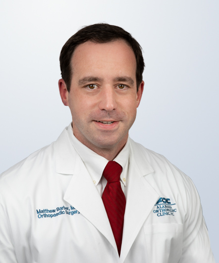
Our Physicians


Specialties


Clinic & Services
We are a full-service orthopaedic clinic. From surgery to therapy – our number one priority is your health, and our knowledgeable staff is here to assist in meeting all of your orthopaedic needs.
View All ServicesPatient Resources
A comprehensive space provides you with everything you need as a patient. You can request an appointment, find paperwork, or pay your bill in one place.
View All Resources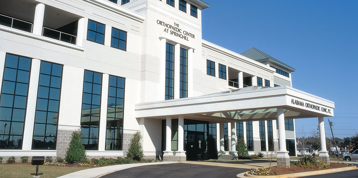
News & Events
News, events, and physician articles – everything AOC and industry-related in one place.
Contact AOC
Our physicians are on call 24 hours a day, seven days a week to handle all of your orthopaedic, pain management, and physical medicine and rehabilitation needs. Correspondence received via the online form is monitored during normal business hours – Monday through Friday, 8am until 5pm. If you have an urgent question after office hours, please call (251) 410-3600.
Phone: 251-410-3600 | Toll Free: 888-878-1999 Fax: 251-410-3700
Office Hours: Monday–Friday 8:00 am–5:00 pm
News & Events

Knee Arthroscopy and Meniscal Tears – Part One
Knee arthroscopy (the placing of a small camera into the knee and performing surgery through small holes) is one of the most common orthopaedic procedures performed in the United States. Over 900,000 of these are performed each year, and over half are done to operate on a torn meniscus.1 The meniscus is a C-shaped cartilage structure on the inside (medial) and outside (lateral) of the knee. Acute traumatic tears of the meniscus are often caused by sporting activities or an activity where there is a sudden twisting of the knee. Hyperflexion of the knee can cause meniscal tears also. Degenerative type tears can be caused by age or untreated instability of the knee.
The meniscus serves an important function in the knee by acting as a cushion to the underlying joint cartilage called the articular cartilage. Loss of meniscus tissue through tearing or surgical removal can lead to increases in stress placed upon the articular cartilage. This can lead to arthritis. Additionally, the meniscus acts as a joint stabilizer. Loss of the meniscus can place increased stress on the major ligaments of the knee such as the anterior cruciate ligament (ACL). Therefore, preserving as much meniscus as possible is a goal for orthopaedic surgeons.
The diagnosis of a meniscal tear requires taking a history, performing a physical exam of the entire knee and surrounding structures, and performing additional diagnostic tests. As stated above, a history of a twisting injury or hyperflexion injury of the knee can lead to a meniscal tear. Common physical exam findings include swelling of the knee, joint line tenderness on the side of the tear, and pain with certain maneuvers that the physician performs such as a McMurray’s test. (The McMurray’s test is performed by taking the knee from a flexed position to an extended position while the tibia is kept internally rotated (for the lateral meniscus) or externally rotated (for medial meniscus). McMurray described a palpable click as being “positive”.2
Diagnostic tests for evaluating a torn meniscus include X-rays of the knee, MRI and knee arthroscopy. The plain X-rays do not detect the torn meniscus, but they rule out other causes of knee pain such as loose bodies and arthritis. The most common non-surgical diagnostic test for meniscal tears is the MRI. It has a high sensitivity and does not involve radiation. MRI can detect both meniscal and ligamentous tears. The “gold standard” test for diagnosing meniscal tears is actually seeing it with knee arthroscopy. This common outpatient procedure can be performed under general or spinal anesthesia.
1Kim et al: Increase in outpatient knee arthroscopy in the United States: a comparison of National Surveys of Ambulatory Surgery, 1996 and 2006. J Bone Joint Surg Am. 2011 Jun 1;93(11):994-1000.
2 McMurray TP: The semilunar cartilages. Br J Surg 1942;29(116):407–414
- < Back
- |